What is the Achilles Tendon?
Tendons are the links between the muscles and the bones The tendon is the connection between the muscle and the moving part of the body. It may help to think of the tendon as a rope, and the muscle as a motor. Tendons, unlike muscle which is red, are white. They have a poor blood supply. This poor blood supply means that tendons heal slowly.
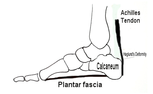
The Achilles tendon is the large tendon that joins the calf muscle to the heel bone (calcaneum). The calf muscle is extremely strong and is responsible for spring and push off during walking and running. The Achilles tendon is the largest tendon in the body and can be felt just above the heel – it is sometimes known as the “the heel cord.”
It is named after the mythical Greek warrior Achilles who, as a child, was dipped into the river Styx by his mother. Submersion made him invulnerable to injury, except for the area of his heel by which he was held. This area remained dry. Achilles was mortally wounded during the siege of Troy when he was struck in the heel by an arrow.
Problems in the Achilles tendon are relatively common as people get older. There are two main diseases affecting the tendon:
- Pain and thickening of the tendon, as a result of degeneration – Achilles tendonosis (or tendinitis).
- Achilles tendon rupture.
Chronic Achilles tendon pain?
Chronic pain and swelling in the Achilles tendon occurs in two locations:
- A lump in the substance of the tendon two to six centimetres above its insertion into the heel bone (non-insertional tendinosis). This pain occurs because of age related changes, which cause the tendon to swell and degenerate. This may be associated with a partial rupture of the tendon. Partial ruptures do not necessarily need surgery, or rest. Sometimes continued stretching is the best option.
- Pain at the point of insertion of the tendon onto the heel bone (insertional tendinitis). This is often associated with a hard, bony lump on the point of the heel. This lump of bone is known as a Haglund’s deformity.
The pain is often most severe on rising in the morning and usually worsens with walking and running. In more severe cases it may occur at rest. Over time, as the symptoms worsen, the pain occurs after less and less activity.
The symptoms may start acutely, with symptoms building up over a couple of weeks. Alternatively it may start gradually.
Investigations
Your doctor may order an X-ray. Often this will show no abnormality but if the tendinitis is lower down at the tendon insertion, the X-ray may show bone spurs. An ultrasound or MRI scan may also be ordered to confirm the diagnosis, and determine how severely the tendon is affected.
What is the Treatment of Achilles Tendonosis?
Without an operation
Most cases of non-insertional Achilles tendonosis are treated successfully without surgery. Usually the longer the symptoms have been present, the longer they will take to settle down. Treatment includes stretching, and regular paracetamol or anti-inflammatory medication.
Physiotherapy is also an important part of the treatment. This includes an intensive program of calf stretches (the eccentric training programme described at the end of this information sheet). Sometimes a medial arch support (or orthosis) may be used. Modifying sporting and training routines can also help. In insertional tendonitis shoes that press less on the heel also help.
Injection of the tendon with steroid tends not to be used as it can weaken the tendon. Occasionally there is a small bursa, or bag of fluid secondary to inflammation, which can be injected with steroid.
With an operation
If non-surgical treatment fails to cure the condition then surgery can be considered. This is more likely to be the case if the pain has been present for six months or more.
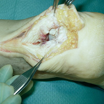
The nature of the surgery depends if you have insertional, or non-insertional disease.
In non-insertional tendonosis the damaged tendon is thinned and cleaned. The damage is then repaired. If there is extensive damage one of the tendons which moves your big toe (the flexor hallucis longus) may be used to reinforce the damaged Achilles tendon.
In insertional tendonosis there is often rubbing of the tendon by a prominent part of the heel bone. This bone is removed. In removing the bone the attachment of the tendon to the bone may be weakened. In these cases the attachment of the tendon to the bone may need to be reinforced with sutures and bone anchors.
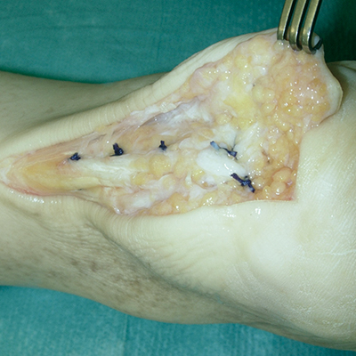
What can I expect after an operation?
For the surgery you will need a general anaesthetic. You will be treated either as a day case, or kept in hospital for one night. Post-operatively it is important to keep the foot elevated. After surgery a plaster splint is usually applied from below the knee to the toes. A prescription for analgesics will be provided. The wound is inspected and the sutures removed after 10 to 14 days. The plaster will be needed for between two and eight weeks, depending on the extent of the surgery to the tendon, and thus the amount of damage present before surgery. Crutches are necessary if you are in plaster. It takes six months for the tendon to fully heal.
Are There Any Risks With Surgery?
The vast majority of patients who have surgery for Achilles tendonosis recover well, without complication. However, as with any surgery there is always a small chance of problems. These include:
- About 85% of patients are satisfied with the results of their surgery, but in some patients the Achilles tendon pain fails to settle despite surgery. In these cases further surgery may be recommended. The surgery is much less successful in smokers.
- It is uncommon for the tendon swelling to resolve fully. But in the absence of pain the swelling is less of a problem.
- Tendon rupture or detachment from the heel bone – this can be repaired, but will require a further operation.
- Infection – this may require intravenous or oral antibiotic treatment.
- Wound healing problems – this is associated with infection and occasionally requires plastic surgery. Wound healing problems are best avoided by keeping the leg elevated after surgery. Smoking increases the risk of this complication.
- Nerve damage – causing pain and numbness in the foot.
- Bleeding.
- Swelling.
The general complications of any surgery and general anaesthesia. Blood clots can occur in the leg (deep venous thrombosis) and in some cases can even go into the lung (pulmonary embolus).
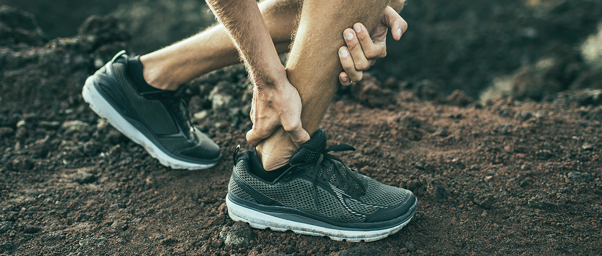
What happens if I rupture my Achilles tendon?
Achilles tendon rupture means a sudden snapping of the tendon. This is happening with increasing frequency as a result of people’s increasing participation in sport. The rupture is spontaneous and typically occurs in the thirties and forties. The tendon is often weakened because of Achilles tendon degeneration (see Achilles tendonosis above); nevertheless the rupture usually occurs without warning. Partial ruptures usually occur in degenerate tendons, and are often treated with stretching (see above under tendonosis).
Often the individual will be playing a sport such as squash, tennis or netball. After a jump or push off there is a sudden snapping sensation in the Achilles tendon area, followed by pain. Sometimes the snap is so loud that it can be heard. The patient often feels that they have been kicked or struck in the heel. This is followed by swelling and bruising. The foot feels weak, with particular weakness pushing off the ground.
How can the rupture be diagnosed by my Doctor?
The normal site of tendon rupture is just above its insertion onto the heel bone. Often a gap can be felt in the tendon and the foot does not move normally when the calf is squeezed. Sometimes an ultrasound or MRI Scan is used to confirm the diagnosis.
What is the Treatment of Achilles tendon ruptures?
It is important that an Achilles tendon rupture is treated adequately; otherwise there will be ongoing weakness. This weakness will interfere with walking and sports activities, and may require later reconstructive surgery. Nevertheless, treatment can be either with or without an operation. Whether you undergo surgery, or not, is a judgement based upon a discussion between you and your surgeon
Without an operation
Non operative treatment involves casting or bracing the foot and ankle with the foot pointed downwards (plantar flexed or in equinus) for approximately 8 weeks. During this time the position of the foot is changed on several occasions.
The advantage of non-operative treatment is that it does not require surgery, reducing the risk of surgical and anaesthetic complications. The disadvantage of non-operative treatment is that the risk of tendon re-rupture at a later date is higher (around 18%). If the tendon re-ruptures surgery will be required. Also, the calf muscle and Achilles tendon may have slightly less power than if the tendon is repaired with an operation.
With an operation
Operative treatment for Achilles tendon rupture involves making a small incision over the site of rupture. The two ends of the tendon are sewn together with stitches. On waking from the operation the foot and ankle is in a plaster splint which holds the foot pointing downwards. Typically you will be in hospital overnight and up on crutches the next day. It is important to keep the foot elevated for the first week or two after surgery to reduce the swelling. You will be on crutches with a splint for 6 to 8 weeks. The position of the splint is changed to bring the foot up to square over that period.
The advantage of surgical treatment is that the chance of the tendon re-rupturing at a later date is lower (around 2%) and the strength and endurance of the calf muscle and Achilles tendon is slightly greater.
Whether the tendon rupture is treated with or without surgery, physiotherapy for strengthening and range of motion may be necessary when the tendon ends have healed together. This strength and range of motion of the foot and ankle will gradually come back over the months afterwards and in general it will be five to six months until there is full recovery and you can return to sport. Gentle running on a flat surface can be undertaken at four months after the rupture.
What happens if the diagnosis Achilles tendon rupture is delayed?
Surprisingly missing a rupture is not that rare, and occurs in 1 in 4 cases. If the diagnosis is delayed the damage can usually be repaired with surgery. Although there is likely to be a little more scarring, the functional result is good. Sometime the two ends of the tendon have tightened up, and cannot be sewn together. In these cases the gap may be bridged by lengthening the tendon, or alternatively by using the tendon to the big toe (the flexor hallucis longus) to bridge the gap.
Are There Any Risks With Surgery?
The vast majority of patients who have surgery for Achilles rupture recover well, without complication. However, as with any surgery there is always a small chance of problems. These include:
- The main reason for operating is to reduce the risk of re-rupture, or failure of the tendon to heal. Surgery reduces this risk from about 1 in 6, to 1 in 50.
- Infection – this may require intravenous or oral antibiotic treatment.
- Wound healing problems – this is occasionally associated with infection and occasionally requires plastic surgery. It is best avoided by keeping the leg elevated after surgery. Smoking increases the risk of wound problems.
- Nerve damage – causing pain and numbness in the foot.
- Bleeding.
- Swelling.
- The general complications of any surgery and general anaesthesia. Blood clots can occur in the leg (deep venous thrombosis) and in some cases can even go into the lung (pulmonary embolus).
Tendo-Achilles Tendinosis – Eccentric Training Programme
The exercises should be supervised and progressed by a registered physiotherapist
Warm-up
- Move ankle up and down X 30.
- Walk backwards with toe/heel pattern for 3 mins.
Wall Knee Squat
- Stand facing close to a wall, feet shoulder width apart. Bend knees so that they lightly touch the wall. Return to the start. Repeat X 10.
- Progress by bending knees towards the wall and left, and then repeat to the right. (e.g. skiing position)
- Progress further by exercising on one foot only.
Balance & Reach Toe/Knee Stretch
- Balance on the injured foot facing the wall approximately an arms length away.
- Stretch to the skirting-board with the un-injured leg, bending the supporting leg.
- Touch the bottom of the skirting-board/floor and return to the stating position, repeat by now stretching on the left side whilst keeping your balance. Return to the start position and repeat process stretching to the right side.
- Repeat whole process X 10
Eccentric Lowering
- Stand on the edge of a step, keep body upright, knees straight and take body weight into good leg, raise up onto toes, then transfer weight onto injured leg and lower down from tip toes back to horizontal.
- Repeat X 10 (h to 15) X 3 sets.
- Progress by –
i. increasing the range of movement by taking the heel below the step level.
ii. increase the load by adding weight e.g. hand weights, multi-gym press or rucksack.
Exercise 4 can be repeated with knees bent.
Activity
Jogging and walking is allowed if it can be performed with only mild discomfort and no pain. Initially start at a slow pace, for a short distance and on flat ground. Gradually increase the activity if there is NO severe pain in the tendon.
No training should be undertaken through disabling pain.